USAID DEC
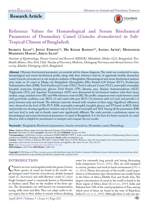
Diversity and prevalence of parasitic infestation with zoonotic potential in dromedary camel (Camelus dromedarius) and fat-tailed sheep (dhumba) in Bangladesh
Sign inHEALTH ALLIANCE INTERNATIONAL
Diversity and prevalence of parasitic infestation with zoonotic potential in dromedary camel (Camelus dromedarius) and fat-tailed sheep (dhumba) in Bangladesh
Dromedary camel (Camelus dromedarius) and fat-tailed sheep (dhumba) are well-known livestock species, primarily reared for meat or milk, skin, and wool.
2019 · 6 pages
Sign in to readA free account is required to download.
Abstract
They are mostly found in desert areas of Africa, the Middle East, and South Asia. In Bangladesh, these species are not common, but they hold high economic value during religious festivals. The only established camel farm in Bangladesh was established in 2006 with six imported camels from India and has since been successful in breeding and increasing the number of animals. Parasitic infestation is a major constraint for profitable livestock production in tropical and sub-tropical countries. Parasites directly or indirectly affect production by reducing fertility and work capacity, decreasing food intake, and lowering weight gain and milk production, and lead to mortality in heavily parasitized animals. Public health is in danger due to zoonotic parasites. Fat-tailed sheep and camel are more resistant to disease than other domesticated livestock, but different types of parasites have been identified in dromedaries and sheep from various countries. A study conducted in Bangladesh reported a gastro-intestinal (GI) parasite prevalence of 27.9%, 26.3%, and 26.2% in 2012, 2013, and 2014, respectively, with zoonotic parasite Fasciola hepatica detected in that study. Previously, F. hepatica had been identified in dromedary, responsible for hydatid disease and fascioliasis in humans. Knowledge about parasitic infestation in dromedary camel and dhumba and their zoonotic significance is scarce in Bangladesh. Therefore, the present study was conducted to provide preliminary information about prevalence and type of GI parasite of dromedary camel and fat-tailed sheep, and hemoparasites of camel. A total of 87 fecal samples (55 camels, 32 dhumba) and 55 camel blood samples were collected during September-October 2015 from a farm and a livestock market in Dhaka. The samples were examined using direct smear, sedimentation technique, flotation technique, and McMaster technique for GI parasite detection. Giemsa stained blood smears were examined under microscope for hemoparasite detection. The overall prevalence of GI parasite was 62% (n = 34; 95% CI: 47.73-74.59) in camel and 59.4% (n = 19; 95% CI: 40.65-76.30) in dhumba. Among the sampled camel, 34 (61.8%; 95% CI: 47.73-74.59) were infected with at least one genus of parasite, with 15% harboring more than one genus of parasite. The prevalence of GI parasite and hemoparasite in camel were recorded as Trichuris spp. (n = 16; 29%; 95% CI: 17.6-42.9), Balantidium coli (n = 12; 22%; 95% CI: 11.8-35.0), Trichostrongylus spp. (n = 7; 13%; 95% CI: 5.3-24.5), Strongyloides spp. (n = 5; 9%; 95% CI: 3.0-20.0), Anaplasma spp. (n = 5; 9%; 95% CI: 3.02-20.0), Paragonimus spp. (n = 1; 2%; 95% CI: 0.05-9.7), Schistosoma spp. (n = 1; 2%; 95% CI: 0.05-9.7), Hymenolepis spp. (n = 1; 2%; 95% CI: 0.05-9.7), Moniezia spp. (n = 1; 2%; 95% CI: 0.05-9.7), and Babesia spp. (n = 1; 2%; 95% CI: 0.05-9.7). The mean EPG feces of camel was 291.76 ± 42.03 with a range of 0-1,400. Total 59.4% dhumba (n = 19; 95% CI: 41-76) were positive for GI parasite, including Trichostrongylus spp. (n = 10; 31.3%; 95% CI: 16.1-50), Strongyloides spp. (n = 9; 28%; 95% CI: 13.8-46.8), B. coli (n = 5; 15.6%; 95% CI: 5.3-32.8), and Trichur